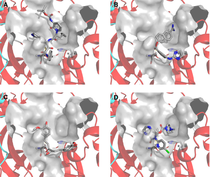
Fig. 2.

The most stable docking mode obtained from autodock vina docking simulations. (A) Binding mode of peptidomimetic α‐ketoamide inhibitor in the crystal structure. (B) Sepimostat, (C) curcumin, and (D) eszopiclone. Chains A and B of the Mpro homodimer are shown as red and cyan ribbons, respectively. The compound, Cys145, and His41 are shown as tubes. Van der Waals surface of the active site is shown in gray color. Water molecules and hydrogen atoms are neglected.
