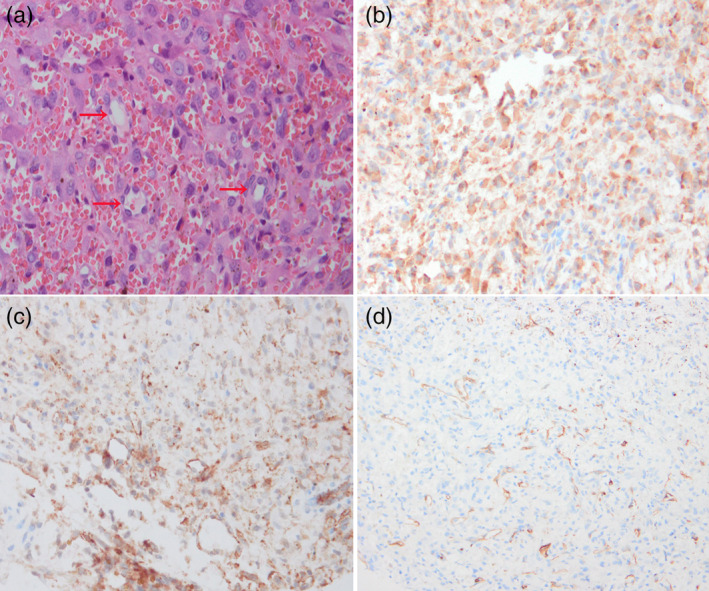
Figure 3.

(a) Hematoxylin and eosin staining showed some dendritic and endothelial cells formed various size of vessels with red blood cells contained (arrows). There was also a dense inflammatory infiltration in some areas. (b–d) Immunohistochemical staining was positive for CAMTA, CD31 and CD34 proteins.
