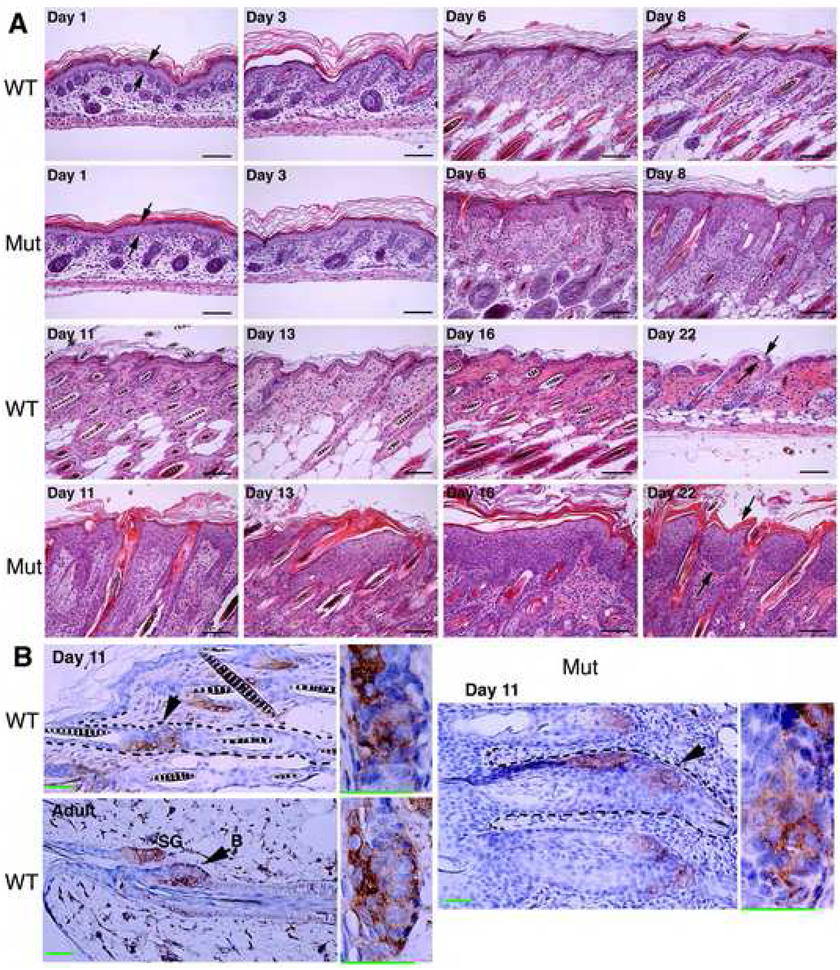
Figure 2. IKKα Deletion Causes Epidermal Hyperplasia.

(A) Histology of the skin of indicated mice, stained with hematoxylin and eosin (H&E). WT, wild-type mice; Mut, IkkαF/F/K5.Cre mice; Day, postnatal day; arrows, indicating epidermis. Scale bars, 30 μm.
(B) Brown, CD34-positive cells indicated by arrows, enlarged in boxes; blue, nuclear counter-staining; SG, sebaceous gland; adult, control for CD34-positive cells in bulge. Scale bars, 30 μm.
