
Figure 6. IKKα Switches Keratinocyte Proliferation and Differentiation via the EGFR Pathway.
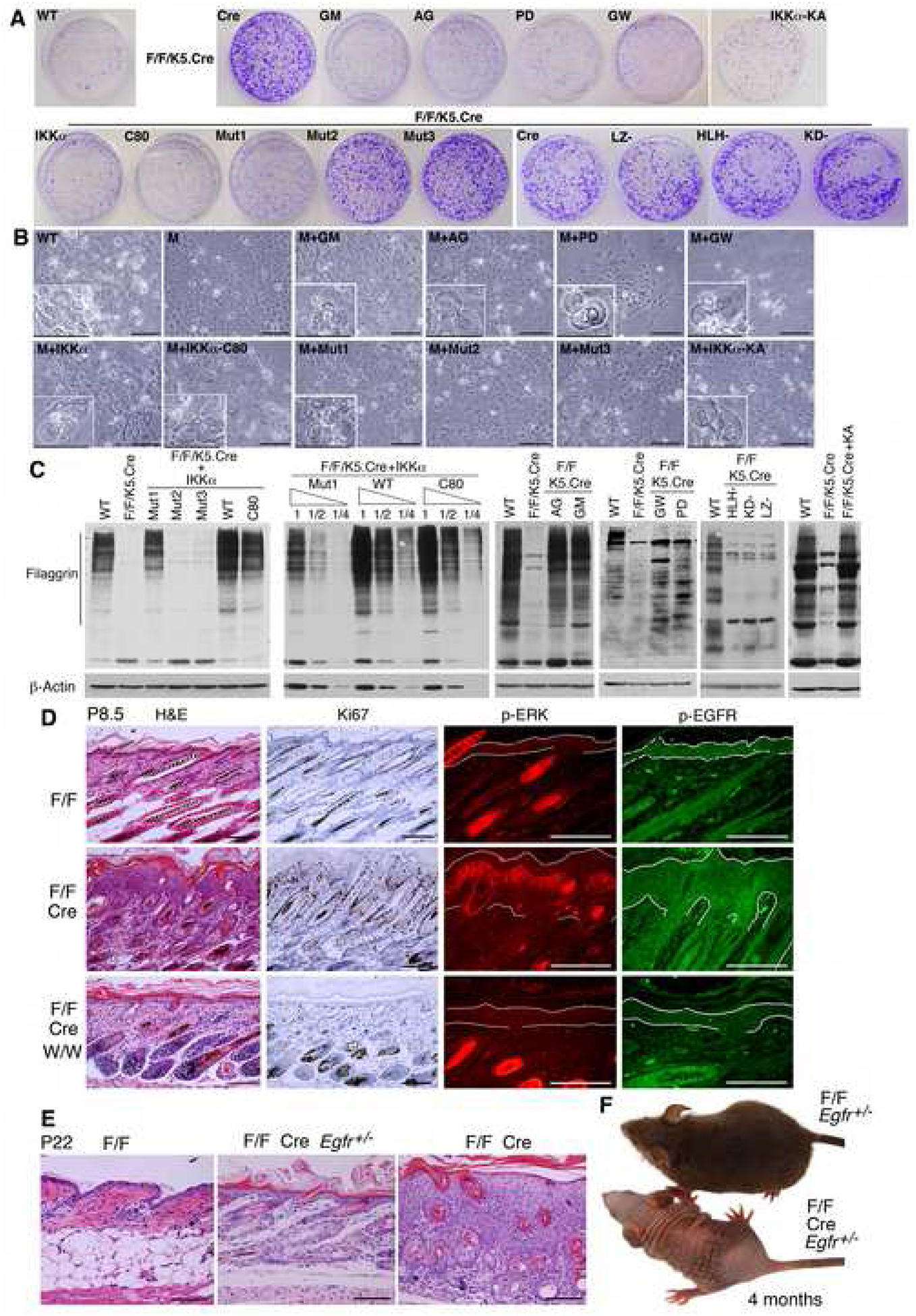
(A) Effects of different forms of IKKα and inhibitors on colony formation of indicated keratinocytes. Different IKKα forms include IKKα, C80, Mut1, Mut2, Mut3, LZ-, HLH-, KD-, and KA. AG, AG1478; GM, GM6001; GW, GW2974; PD, PD98059; keratinocyte colonies, stained with 0.5% crystal violet; WT, IkkαF/F keratinocytes; F/F/K5.Cre, IkkαF/F/K5.Cre keratinocytes.
(B) Keratinocyte morphologies in culture. Cells in small boxes represent terminally differentiated keratinocytes. M, IkkαF/F/K5.Cre keratinocytes. Scale bars, 40 μm.
(C) Filaggrin levels in indicated keratinocytes, as detected by Western blotting. 1, ½, ¼, protein dilutions, β-Actin, loading controls.
(D) The effect of inactivation of EGFR on the epidermis in the indicated mice. P, postnatal day; W/W, Egfrwa2/wa2; Cre, K5.Cre; H&E, hematoxylin and eosin; Ki67, dark brown color stained by immunohistochemistry; p-ERK, red color stained by immunofluorescence; p-EGFR, green stained by immunofluorescence; lines in p-ERK and p-EGFR, limits of the epidermis. Scale bars, 50 μm.
(E) Skin histology of indicated mice, stained with H&E. Scale bars, 60 μm.
(F) Appearances of indicated mice at 4 months of age.