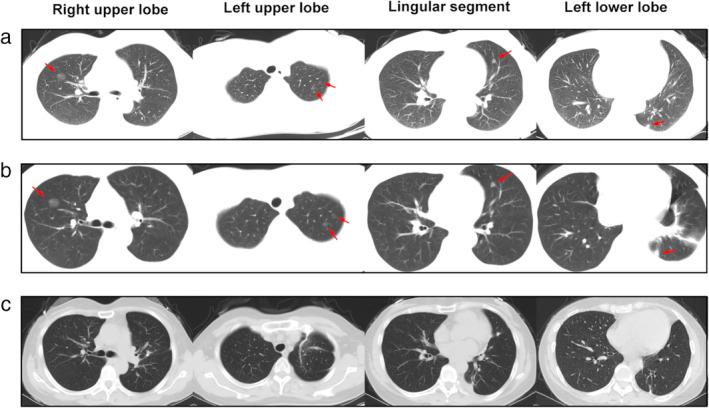
Figure 1.

(a) CT image after trauma showed a pure ground‐glass nodule (pGGN) of approximately 14 mm in the right upper lobe, two pGGNs of approximately 6 mm in the apical segment of the left upper lobe, a mixed GGN (mGGN) of approximately 8 mm in the left lingular segment, and a mGGN of approximately 8 mm in the left lower lobe. (b) There were no significant changes in the nodules after one week of anti‐inflammatory treatment. (c) The pGGN in right upper lobe had disappeared. Shadows of staples and postoperative changes can be seen in the left upper and lower lobes.
