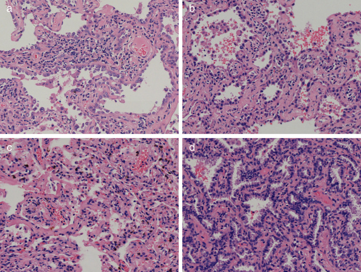
Figure 3.

Pathology of the pulmonary nodules (HE stain, 20×). (a) The nodule in the right upper lobe was adenocarcinoma in situ (AIS). (b) The two nodules in the left upper lobe were AIS. (c) The nodule in the lingular segment was chronic inflammation. (d) The nodule in the left lower lobe was confirmed to be minimally invasive adenocarcinoma (MIA).
