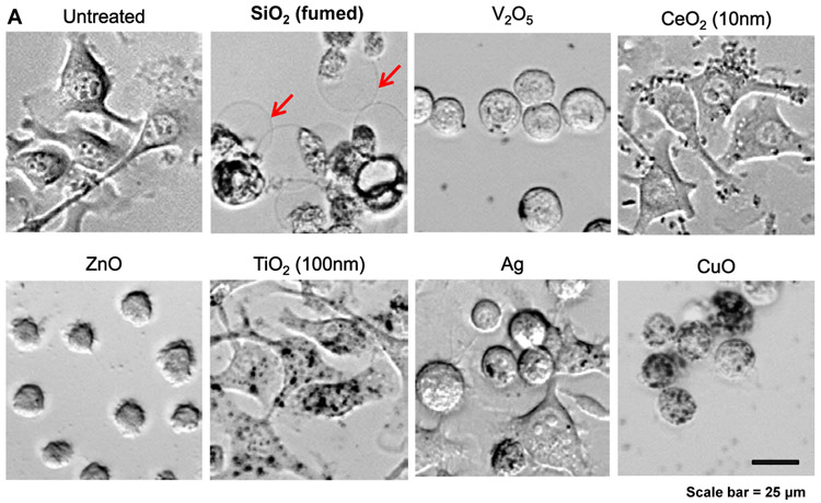
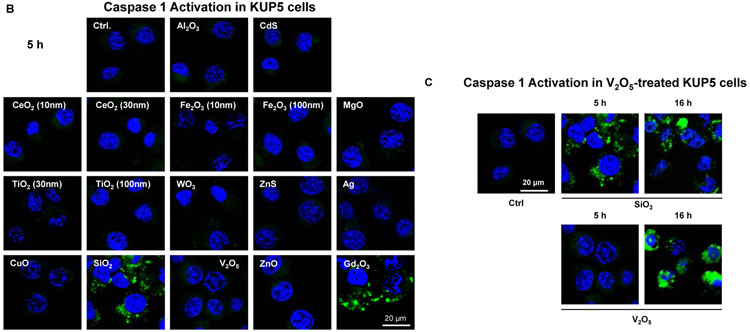
Figure 4. Assessment of nanoparticles to induce Caspase 1 and SiO2 induced pyroptotic cell death in KUP5 cells.
(A) Optical microscopy images of ENM-treated KUP5 cells. Optical microscopy images showing the morphology of KUP5 cells exposed to seven of the NPs (fumed SiO2, Ag, CeO2, CuO, V2O5, TiO2 and ZnO) at 12.5 μg/mL for 6 h. The scale bar is 25 μm. The rest of the optical images for other particles appear in Figure S3. (B) Assessment of caspase 1 activation in ENM-treated KUP5 cells. The LPS-primed KUP5 cells were incubated with nanoparticles at 25 μg/mL for 5 h. Cells were washed with PBS and stained with FAM-FLICA caspase substrate for 1 h at 37 °C. Cells were then stained with Hoechst 33342. (C) Time-dependent activation of caspase 1 in V2O5-treated KUP5 cells. The LPS-primed KUP5 cells were incubated with fumed SiO2 and V2O5 for 5 and 16 h, and the staining process was repeated. The cells were imaged using Leica Confocal SP8-SMD microscope. The scale bar is 20 μm.


