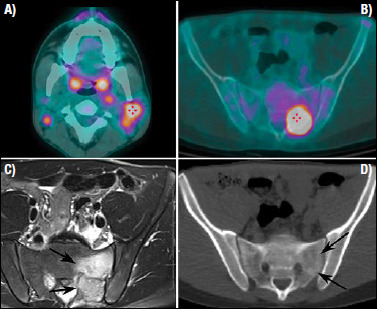
Figure 4.4.

Nasopharyngeal undifferentiated carcinoma in a young patient. (A) The FDG-PET/CT shows a metabolic active adenopathy at level 2b, on the left side of the neck, and a large bone metastases (B) in the left ala of the sacrum, both showing a high metabolic activity. While the CT image obtained during the PET/CT study does not demonstrate sufficient changes of the bony architecture (D), the MRI examination clearly defines the metastasis replacing the posterior aspect of the ala of the sacrum (arrows) and the peritumoural bone oedema (C).
