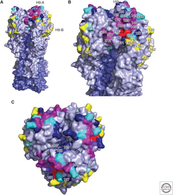
Figure 4.
Relevant structural features of the hemagglutinin (HA) of the H9 subtype. Homotrimers of the HA crystal structure of A/swine/Hong Kong/9/1998 (Protein databank ID:1JSD) colored in PyMOL. Selected receptor-binding site (RBS) residues are colored in red. HA1 and HA2 portions are highlighted in gray and slate blue, respectively. (A) The full HA homotrimer is shown. (B,C) Details of the HA globular head. Shown are the antigenic site H9-A (magenta) and H9-B (yellow). Other antigenic residues without assigned site classification are shown in cyan. Potential glycosylation sites are colored in dark blue.

