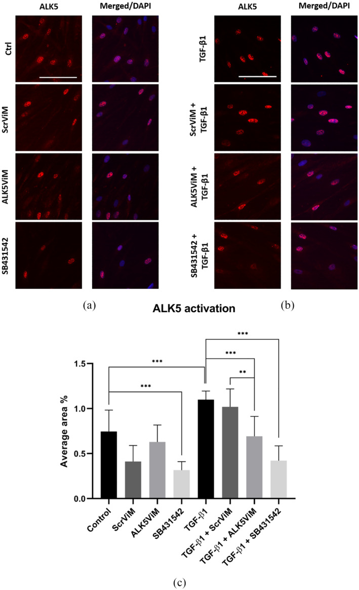
Figure 5.
Immunofluorescence and quantification of activated ALK5 in HS-derived fibroblasts. Fibroblasts were pre-treated with ViMs (2 μM) or SB431542 (10 μM) for 24 h. Next, fibroblasts were treated with hTGF-β1 (5 ng/mL) for 1 h. (a) ALK5 IF staining (red) for the following conditions: TGF-β1, ScrViM+TGF-β1, ALK5ViM+TGF-β1 and SB431542+TGF-β1. Nuclear staining was performed with DAPI, displayed in corresponding merged images. Scale bar: 100 µm. (b) Quantification of ALK5 activation presented as average area fraction. Fluorescent signal was calculated as area fraction for every donor in eight different conditions (Control [untreated], ScrViM, ALK5ViM, SB431542, TGF-β1, ScrViM+TGF-β1, ALK5ViM+TGF-β1 and SB431542+TGF-β1) and was calculated with ImageJ. n = 3; *P < 0.05; **P < 0.01; ***P < 0.001; one-way ANOVA, error bars indicate standard error of the mean.

