Figure 5.
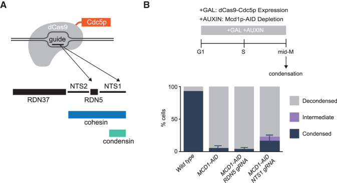
Localization of Cdc5p with CRISPR–Cas9 to the condensin-bound RDN partially restores the RDN condensation defect of cohesin-depleted cells. (A) Galactose addition to the media drives the expression of a catalytically dead dCas9 fused to Cdc5p. Guide RNAs recruit dCas9-Cdc5p fusion protein either to the condensin-bound region NTS1 (RDN5 gRNA) or 500 bp to the left at RDN5 (RDN5 gRNA) where only cohesin is bound. (B, top) “Staged mid-M” regimen used to assess condensation in mid-M. Cultures were synchronized in G1 and released in media containing galactose and auxin to drive the expression of dCas9-Cdc5p concomitantly to the depletion of Mcd1p-AID. Media also contained nocodazole to arrest cells in mid-M and assess condensation. (Bottom) Wild-type, Mcd1p-depleted, Mcd1p-depleted with dCas9-Cdc5p and RDN5 gRNA-expressing, and Mcd1p-depleted with dCas9-Cdc5p and NTS1 gRNA-expressing cells were treated with “staged mid-M” regimen and processed to score RDN condensation. Average of three experiments scoring 100 cells are shown and error bars represent the standard deviation.

