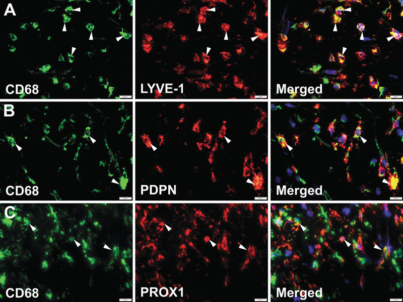
Fig. 7.1.

Human clinical breast cancers massively recruit M-LECP. Human BC specimens were co-stained for CD68 (green) and antibodies against markers of lymphatic vessels (red) including (a) LYVE-1, (b) PDPN, and (c) PROX1. Nuclei in merged images were identified by Hoechst stain. White arrowheads indicate cells that co-express CD68 and lymphatic markers. All images were acquired at 600× magnification
