. 2020 Apr 14;295(3):616–625. doi: 10.1148/radiol.2020192210

Figure 2b:

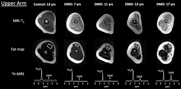
Example upper extremity MRI and proton (1H) MR spectroscopy (MRS) data acquired from the dominant (a) shoulder and (b) upper arm in control participants and participants with Duchenne muscular dystrophy (DMD) at different ages. Single voxel stimulated-echo acquisition mode 1H MR spectroscopy spectra were acquired at echo time of 27 msec from the deltoid (a) and biceps brachii (b).

Example upper extremity MRI and proton (1H) MR spectroscopy (MRS) data acquired from the dominant (a) shoulder and (b) upper arm in control participants and participants with Duchenne muscular dystrophy (DMD) at different ages. Single voxel stimulated-echo acquisition mode 1H MR spectroscopy spectra were acquired at echo time of 27 msec from the deltoid (a) and biceps brachii (b).