. 2020 Apr 7;295(3):583–590. doi: 10.1148/radiol.2020191016

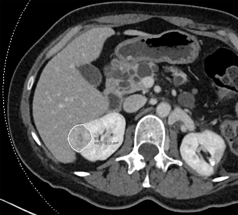
Figure 2b:

Images of clear cell renal cell carcinoma in a 55-year-old man with Von Hippel–Lindau syndrome. (a, b) Axial contrast material–enhanced nephrographic phase CT scans show the tumor was 7.4 cm3 at baseline (a) and had grown to 9.1 cm3 at 125 days (b). (c) Axial apparent diffusion coefficient (ADC) map of the tumor at baseline and (d) corresponding histogram percentile analysis.

Images of clear cell renal cell carcinoma in a 55-year-old man with Von Hippel–Lindau syndrome. (a, b) Axial contrast material–enhanced nephrographic phase CT scans show the tumor was 7.4 cm3 at baseline (a) and had grown to 9.1 cm3 at 125 days (b). (c) Axial apparent diffusion coefficient (ADC) map of the tumor at baseline and (d) corresponding histogram percentile analysis.