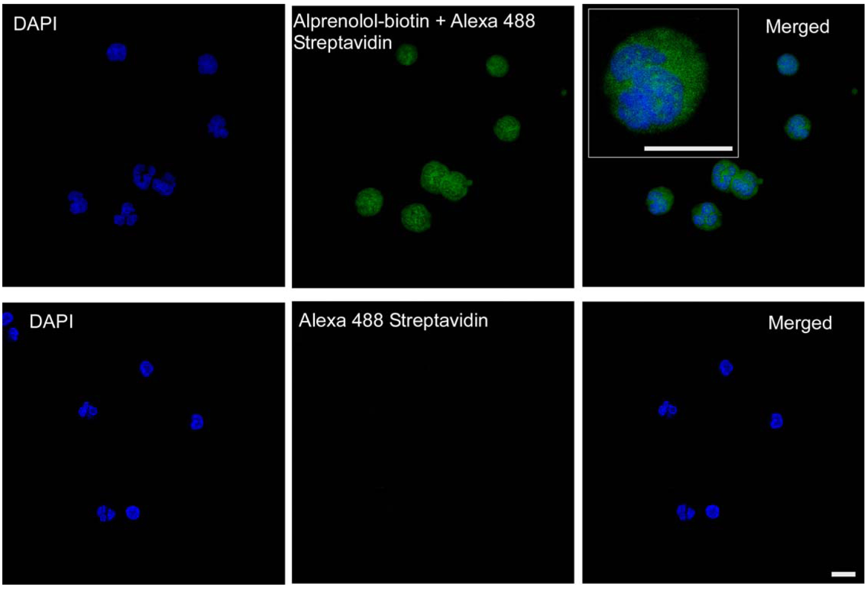
Figure 2.

Imaging of Alprenolol Probe Binding. Confocal microscopy was used to image peneration and intracellular binding of the alprenolol probe. Upper panels show images obtained from DAPI and alprenolol probe channels and both channels merged. Inset shows a high-power image of a cell. Lower panels show staining with DAPI and secondary reagent only. Scale bar = 10 μm.
