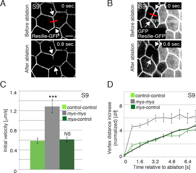
Fig 3. Loss of integrins in FCs results in increased membrane tension.
(A, B) Images of life S9 wild type (A) and mosaic egg chambers containing mys FC clones (GFP-negative) (B), expressing Resille-GFP, before and after single-cell bonds are ablated. Red bar and arrows indicate the point of ablation and the vertexes displaced, respectively. (C) Quantification of initial velocity of vertex displacement and (D) vertex displacement over time of the indicated ablated bonds. The statistical significance of differences was assessed with a t-test, *** P value < 0.0001. All error bars indicate s. e. Scale bars, 5 μm.

