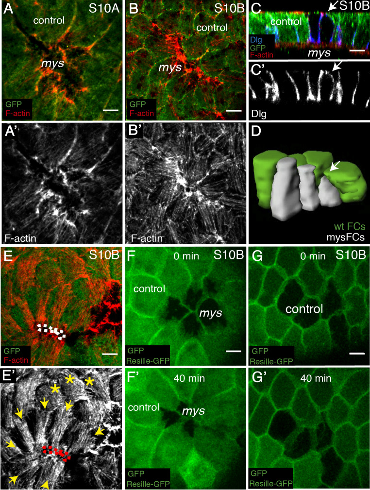
Fig 6. Elimination of integrin in FCs disrupts cytoskeletal organisation in neighbouring control cells.
(A, B) Basal surface view of S10A (A, A’) and S10B (B, B’) mosaic follicular epithelia containing mys FC clones (GFP-negative), stained with anti-GFP (green) and Rhodamine Phalloidin to detect F-actin (red). (C) Lateral view of a S10 mosaic egg chamber stained with anti-GFP (green), Rhodamine Phalloidin (red) and anti-Dlg (basolateral polarity marker Discs large, blue). (D) 3D reconstruction of mys FCs and surrounding control cells. Arrows in C and D point to the basal surface of a mutant FC. (E, E’) Basal surface view of a S10B mosaic FE containing mys FC clones (GFP-negative). Yellow arrows and asterisks mark control FCs contacting control and mys FCs, respectively. (F-G’) Confocal images of live S10B mosaic egg chambers containing mys (F) or GFP (G) clones and expressing the cell membrane marker Resille-GFP. Images were taken with a 40 minutes interval. Scale bars, 5μm.

