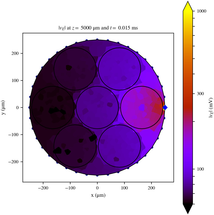
Fig 1. Cross-sectional slice of the extracellular field generated by the electrode.
Cross-sectional slice of the extracellular field generated by the electrode over the model Nerve 1 at the middle of its length (z = 5000 μm), where the stimulation pad (blue diamond) is situated, and at the time step following the onset of the stimulating pulse. The RN assumes the field is constant over the surface of each tessellation polygon. The contours of the nerve and the fascicles are indicated with a black solid line for better identification. Axons are not shown in this figure. Although the maximum value of |vE|, situated at the active site, is 2413.62 mV, the colorbar was cut at 1000 mV in order to facilitate the visualisation of the spatial details of the field.

