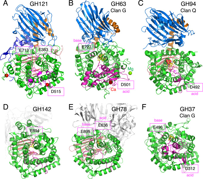
Fig 5. Structural comparison of HypBA2 with structural homologs belonging to other GH families.
Overall structures of (A) GH121 HypBA2, (B) GH63 α-glycosidase YgjK from E. coli (PDB ID 5CA3), (C) GH94 chitobiose phosphorylase ChBP from V. proteolyticus (1V7X), (D) GH142 β-L-arabinofuranosidase BT_1020 from B. thetaiotaomicron (5MQS), (E) GH78 α-L-rhamnosidase SaRha78A from S. avermitilis (3W5N), and (F) GH37 α,α-trehalase Tre37A from E. coli (2JF4) are shown. Catalytic residues are indicated with a magenta color. The A’-region (insertion between the fifth and sixth helices of the barrel) is also shown with a magenta color.

