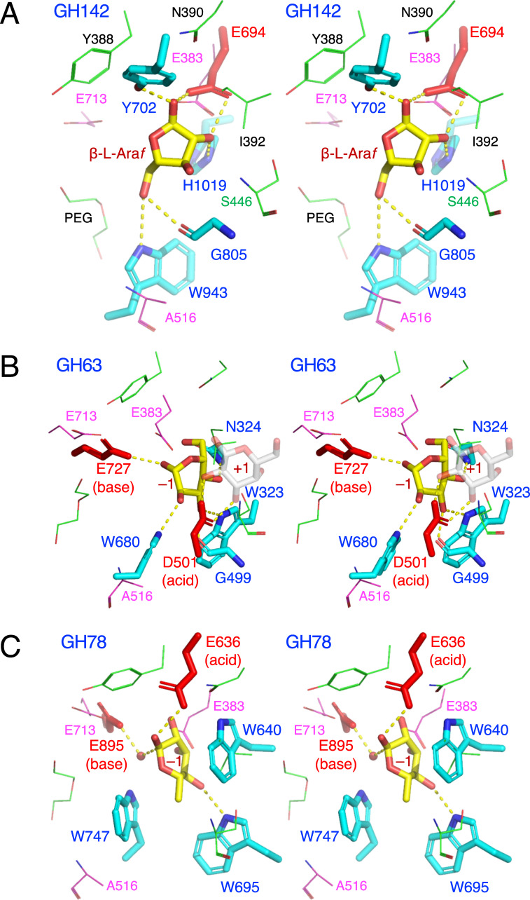
Fig 6. Stereoview of the active site of structural homologs belonging to other GH families.
(A) GH142 β-L-arabinofuranosidase BT_1020 (cyan) complexed with β-L-Araf (yellow), (B) GH63 α-glycosidase YgjK (cyan) complexed with glucose (yellow) and lactose (galactose at subsite +1 is shown as transparent grey sticks), and (C) GH78 α-L-rhamnosidase SaRha78A (cyan) complexed with α-L-rhamnose (yellow). The catalytic residues of BT_1020, YgjK, and SaRha78A are shown as red sticks. Residues in the active site of HypBA2 are superimposed as magenta (putative catalytic residues) or thin green lines.

