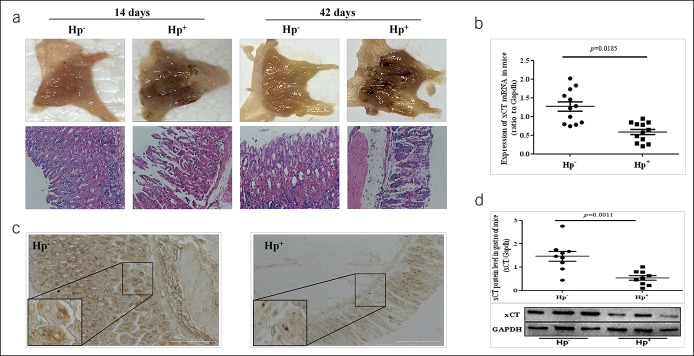
Figure 1.
Decreased cystine-glutamate transporter (xCT) activity in gastric ulcer of H. pylori-infected mice. (a) Photographs of gastric mucosal lesions showed sporadic hemorrhagic spots in gastric mucosa. Hematoxylin-eosin staining showed gastric mucosal damage with dilation and exfoliation of gastric epithelial cells and disruption of mucosal in H. pylori -infected mice with an infiltration of inflammatory cells in the mucosa and submucosa. (b) Decreased expression of xCT mRNA in Hp+ group was detected. (c) Immunohistochemical staining for xCT antigen in gastric tissue. (d) Western blot for xCT in gastric tissue. Data are means ± SEM, N = 8–12, *P < 0.05 vs Hp−, **P < 0.01 vs Hp−. GAPDH, glyceraldehyde phosphate dehydrogenase; Hp, Helicobacter pylori; mRNA, messenger RNA.

