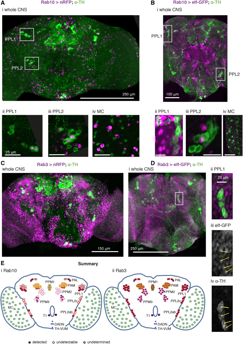
Figure 6.
Rab10 and Rab3 are located in different subsets of the dopaminergic neurons. A, B. Rab10 is detected in some of the dopaminergic neurons that control vision (PPL1, Aii, Bii; PPL2 Aiii, Biii). Not all dopaminergic neurons, identified by a cytosolic α-Tyrosine Hydroxylase antibody (α-TH, green), are indicated by Rab10-GAL4 expression of a strong nuclear RFP or the mainly nuclear eIf-GFP (magenta). The dopaminergic MC neurons in the visual lobes do not stain well with fluorescent reporters (Nassel and Elekes 1992; Hindle et al. 2013) and we could not detect Rab10-driven fluorescence (MC, Aiv, Biv, marked with gray in E). C, D. Rab 3 is present in all dopaminergic neurons. Rab3-GAL4 driven nuclear RFP or eIf-GFP (magenta) marks most neurons, including nearly all that are dopaminergic (green). The PPL neurons not marked by Rab10 expression are included (Dii-iv). E. Summary of the expression pattern of (i) Rab10 and (ii) Rab3. The MC neurons in the optic lobe (Nassel et al. 1988) are also called Mi15 neurons (Davis et al. 2020). Ai, Bi, Ci and Di: projection of confocal stacks through the whole CNS; Aii, Aiii, Bii, Biii, Dii-iv projections of confocal stacks through the cell groups, approximately marked in the whole CNS image; Aiv and Biv sections from a separate preparation to Ai and Bi. Data representative of at least nine brains (from at least 3 crosses), 3-7 days old. The Rab3 > nRFP flies were raised at 18 °C to improve viability. Exact genotypes: +; RedStinger4 nRFP/+; Rab10 Gal4/+; or +; RedStinger4 nRFP/+; Rab3 Gal4/+; or +; eIf-4A3-GFP/+; Rab10 Gal4/+; or +; eIf-4A3-GFP/+; Rab3 Gal4/+;

