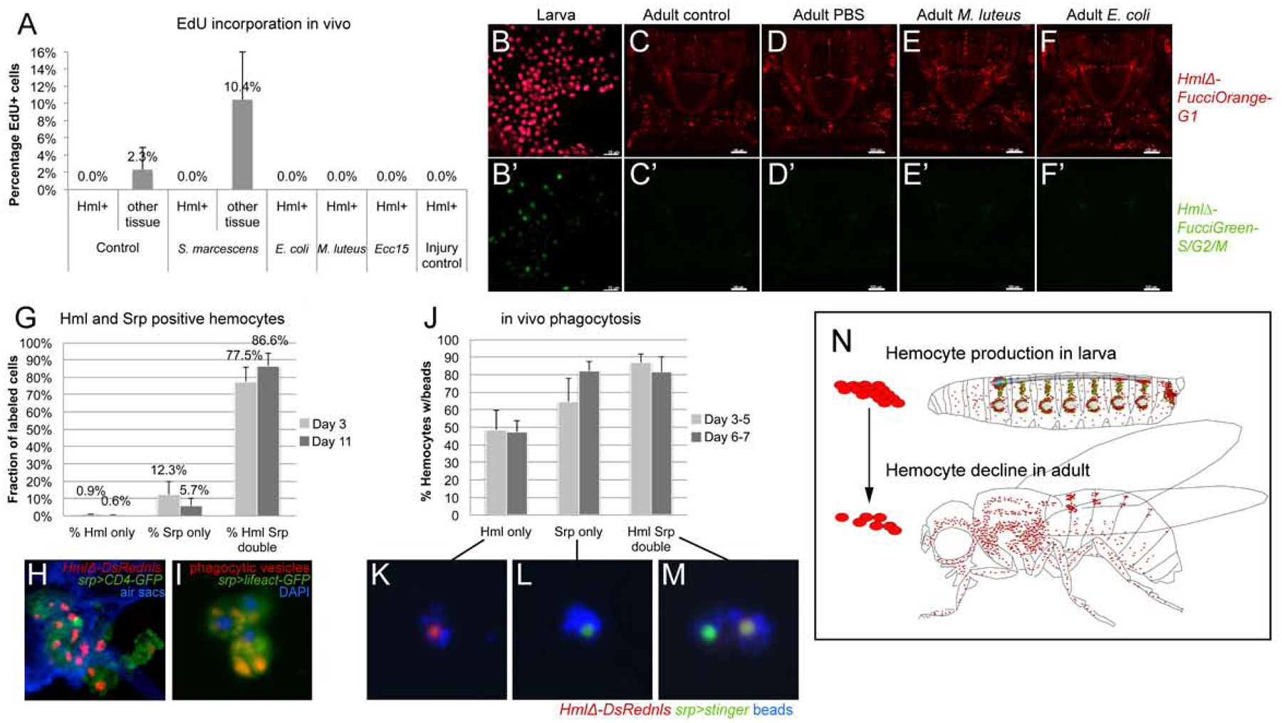
Figure 5. Adult hemocytes do not expand; Srp marks active phagocytes in adult Drosophila.

(A) In vivo EdU incorporation. Percentage of EdU positive cells among hemocytes or control tissue in absence or presence of immune challenges as indicated; average and standard deviation. (B-F’) 2-color Fucci analysis of hemocytes, control (uninfected), sterile injury (PBS), and infection (M. luteus, E. coli); genotype is w1118; HmlΔFucciOrangeG1; HmlΔFucciGreenG2/S/M; (B-B’) embryonic-lineage hemocytes released from larvae, note green cells; (C-F’) imaging of Fucci hemocytes in adult flies, dorsal views of thorax and anterior abdomen, anterior up; note absence of green signal of HmlΔFucciGreenG2/S/M. (G-I) Srp labels phagocytic plasmatocytes in the adult fly. (G) Srp and Hml positive hemocytes, young (3 days) and mature (11 days) adults.. (H) Thorax cross section of adult fly, genotype is HmlΔDsrednls/UAS-CD4-GFP; +/srpD-GAL4; respiratory epithelia (air sacs, blue). (I) Srp-Gal4, UAS-lifeact-GFP positive plasmatocytes (green) with red phagocytic vesicles, released ex vivo from adult fly; DAPI (blue). (J-M) In vivo phagocytosis assay, ex vivo examination of hemocytes. (J) % of hemocytes carrying blue beads, from young (3 days) and mature (11 days) adults. Genotype is HmlΔDsred/ UAS-stinger; +/ srpD-GAL4. (K, L, M) examples of labeled hemocytes as indicated. (N) Model, hemocyte production takes place during the larval stage (mainly in the hematopoietic pockets and the lymph gland), while in the adult hemocyte numbers decline over time.
