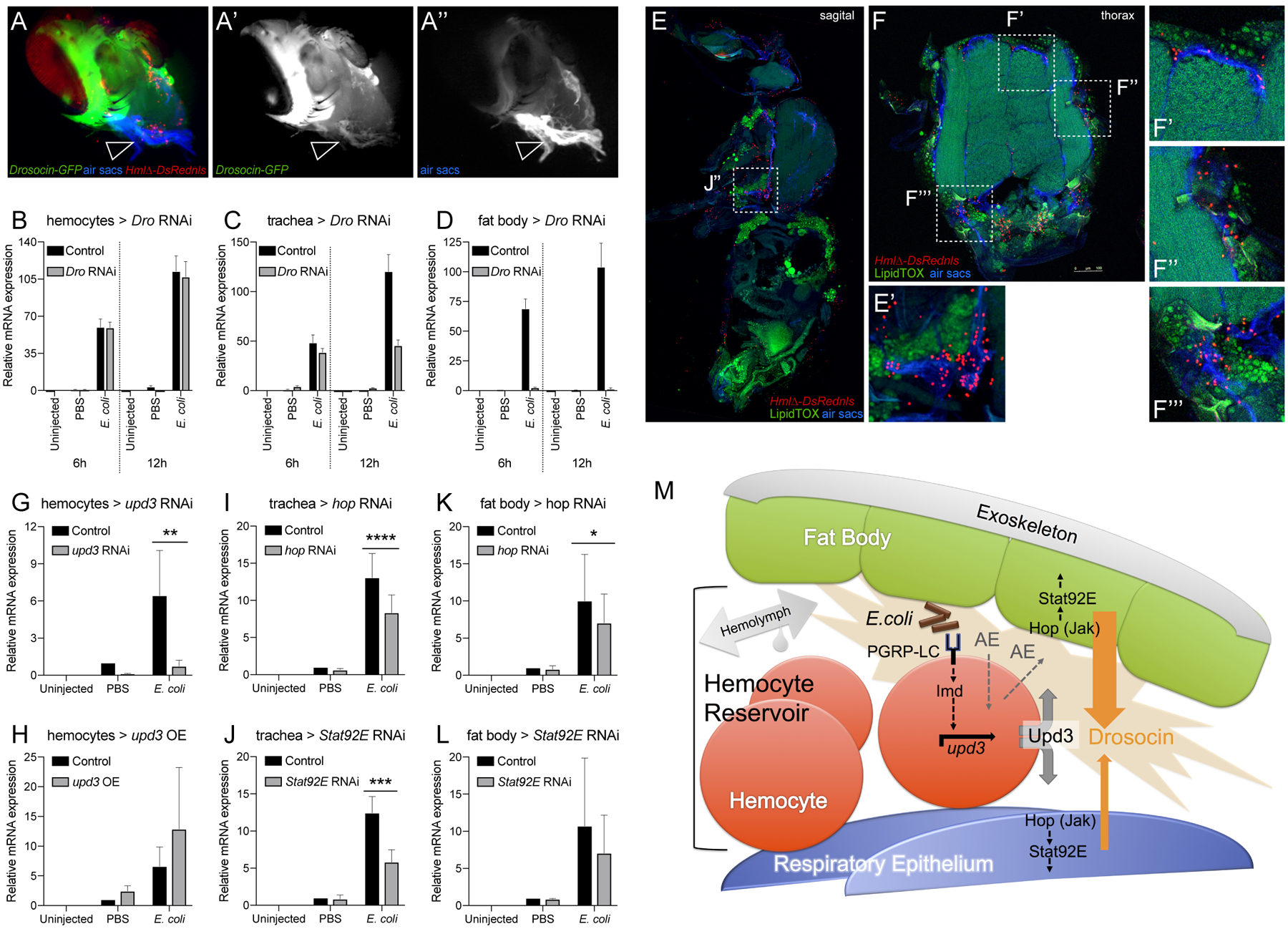
Figure 7. The Drosocin response is localized to the reservoir of hemocytes at the respiratory epithelia and colocalizing fat body domains, and requires Upd3 signaling from hemocytes.

(A-A”) Dissected heads of genotype Drosocin-GFP/HmlΔ-DsRed (Drosocin-GFP green, hemocytes red), respiratory epithelia (air sacs, blue); (A’) Drosocin-GFP, white; (A”) respiratory epithelia, white. Note Drosocin-GFP expression is high in fat body and moderate in respiratory epithelia (arrowhead). (B-D) Tissue specific RNAi knockdown of Drosocin; overall Drosocin mRNA levels were quantified by qPCR. 6–7 day-old adult females were left untreated, injected with PBS, or E.coli in PBS (OD 6), 9.2 nl, and harvested 6 and 12h post infection. Charts display mean and SEM of samples from a representative biological replicate experiment, using pools of 10 females per condition, and triplicate qPCR runs. Values of all charts are displayed relative to the RNA level induced by the sterile PBS injections in control flies. (B) Drosocin RNAi silencing in hemocytes; (C) in respiratory system; (D) in fat body. (E-F’’’) Anatomy of fat body tissue lining the respiratory epithelia and hemocytes; HmlΔ-DsRednls (hemocytes, red), fat body (LipidTOX, large, distinct green cells), respiratory epithelia (air sacs, blue) (E) Sagital section of adult Drosophila. (E’) Closeup of region indicated in (E). (F) Thorax cross section. (F’-F’’’) Closeup of regions indicated in (F). (G-L) Expression of Drosocin in adult flies upon silencing or overexpression of upd3 and silencing of genes of the Jak/Stat pathway. 5 day-old adult Drosophila untreated, injected with sterile PBS, or E.coli in PBS (OD 6), 9.2 nl; flies harvested at 6h post injection. Charts display mean and CI of samples from 3 averaged biological replicate experiments, using pools of 10 females per condition, and triplicate qPCR runs for each sample. Values of all charts are displayed relative to the average RNA level induced by the sterile PBS injections in control flies. Two-way ANOVA with Sidak’s multiple comparison test, *,**,***,or **** corresponding to p≤0.05, 0.01, 0.001, or 0.0001, respectively. (G, H) Drosocin qPCR of whole flies, inducible transgene expression in hemocytes, (G) Genotypes are control (HmlΔ-GAL4,UAS-GFP/+; tub-GAL80ts /+) versus HmlΔ-GAL4,UAS-GFP/+; tub-GAL80ts /UAS-upd3 RNAi. (H) Control versus HmlΔ-GAL4,UAS-GFP/ UAS-upd3; tub-GAL80ts /+. (I, J) Drosocin qPCR of whole flies, inducible transgene expression in tracheal system. (I) Genotypes are control (btl-GAL4, tub-GAL80ts, UAS-GFP /+) versus btl-GAL4, tub-GAL80ts, UAS-GFP / UAS-hop RNAi; (J) Genotypes are control versus btl-GAL4, tub-GAL80ts, UAS-GFP / UAS-Stat92E RNAi. (K, L) Drosocin qPCR of whole flies, transgene expression in fat body. (K) Genotypes are control (ppl-GAL4, UAS-GFP / +) versus ppl-GAL4, UAS-GFP /+; UAS-hop RNAi/+; (L) Genotypes are control versus ppl-GAL4, UAS-GFP/+; UAS-Stat92E RNAi/+. (M) Model of communication between hemocytes, fat body and respiratory epithelia, in which hemocytes act as sentinels of infection. Gram-negative bacteria that accumulate together with hemocytes in the reservoir between respiratory epithelia and fat body; activation of Imd signaling through PGRP-LC on hemocytes triggers upd3 expression and Upd3 secretion. Upd3 activates Jak/Stat signaling in adjacent domains of the fat body and the respiratory epithelia, contributing directly or indirectly to the induction of Drosocin expression. Since we PGRP-LC/Imd signaling and Upd3/Jak/Stat signaling are required but not sufficient to induce Drosocin expression, additional events (AE) in parallel to these pathways are proposed that would provide sufficiency to trigger Drosocin induction.
