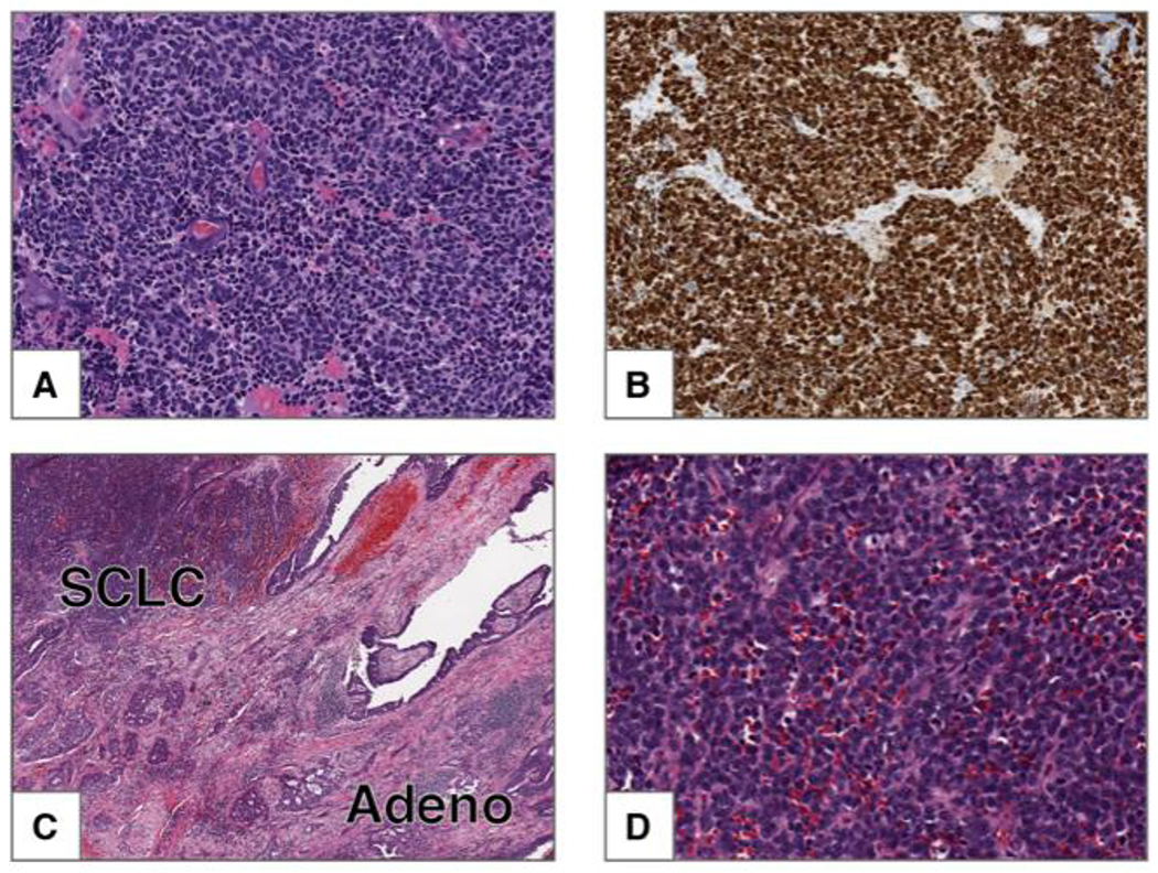
Figure 2: Small cell carcinoma.

A) This tumor is composed of small cells with scant cytoplasm, finely granular chromatin and frequent mitoses. Nucleoli are absent. B) Ki-67 shows strong nuclear staining in 100% of the tumor cells. Combined small cell carcinoma and adenocarcinoma. C) Low power image of tumor composed of two components: small cell carcinoma (upper left) and adenocarcinoma with acinar pattern (lower right). D) High power image of the SCLC component from C.
