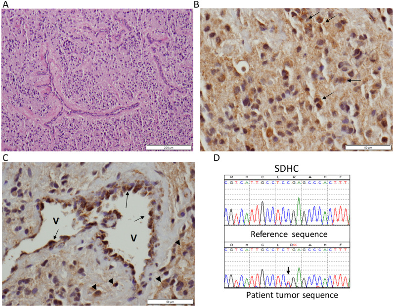
Figure 1.
(A) Hematoxylin and eosin–stained photograph showing the paraganglioma with admixed thin-walled vessels. (B) High magnification of a representative area of the paraganglioma following SDHB immunohistochemistry (IHC): most tumor cells in the field show diffuse weak staining, and rare tumor cells have the more typical “granular” signal of SDHB staining (arrows). (C) Another high-magnification field of SDHB IHC showing negative or weak diffuse staining in the tumor, with a few scattered cells with a stronger, positive granular signal (arrowheads). Note strongly positive cells with granular appearance lining a vessel (V) wall (arrows). Original scale bars are shown at right bottom corners. (D) Sequence trace of the patient’s paraganglioma DNA (lower) showing the SDHC mutation c.43C>CT, p.R15X (arrow). There is no clear evidence of loss of heterozygosity in the tumor.

