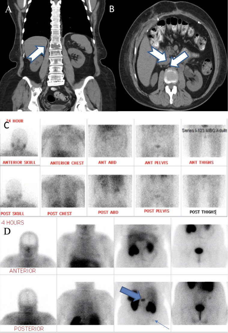
Figure 2.
(A) Coronal view of CT abdomen (ABD) and pelvis without contrast revealing first lumbar vertebral lytic bone lesion (arrow). (B) Transverse view of the same CT scan revealing extensive retroperitoneal lymphadenopathy (arrows). (C) Negative iodine-123-meta-iodobenzylguanidine (I-123 MIBG) scan despite clinical and biochemical evidence of recurrent paraganglioma. (D) Octreotide scan revealing foci of increased radiotracer activity within the first lumbar vertebral body (thick arrow) and faintly at right iliac crest (thin arrow; 300 pixels per inch). ANT, anterior; POST, posterior.

