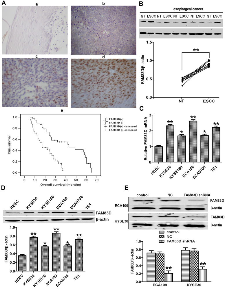
Figure 1.
Expression of FAM83D in human esophageal clinical specimens and cell lines. (A-a–d) Immunohistochemical analysis of FAM83D expression in normal adult esophageal tissues (a) and esophageal carcinoma tissues (b–d). FAM83D staining is mainly localized in the nuclei of cancerous cells and seldomly observed in the cytoplasm and plasmalemma. (a) Weak or absent FAM83D staining in the normal esophageal epithelium. (b–d) Weak, moderate and strong FAM83D staining in esophageal carcinoma. Magnification: ×200. (A-e) Kaplan–Meier overall survival curves for 69 patients with esophageal carcinoma. Compared to low FAM83D expression, high FAM83D expression was associated with a significantly shorter overall survival (P<0.001, Log rank test). (B) Representative Western blotting showing FAM83D expression in 10 pairs of ESCC and nontumor (NT) tissues and comparison of the expression levels of FAM83D protein in different esophageal tissue samples. **P<0.01 compared with NT tissues. (C) Relative mRNA expression of FAM83D in five esophageal cancer cell lines and the normal human esophageal epithelial cell line HEEC. GAPDH was used as a loading control. *P<0.05, **P<0.01 compared with HEECs. (D) Similar evaluation using Western blotting. β-actin was used as a loading control. *P<0.05, **P<0.01 compared with HEECs. (E) Western blotting showed that the expression of FAM83D protein in ECA109 and KYSE30 cells was significantly downregulated by shRNA targeting FAM83D. **P<0.01 compared with the control group and NC group.

