Figure 3.
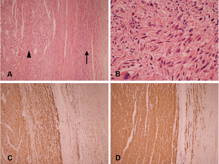
Microscopic details of the tumor. (A) The interlacing bundle and fascicles of the tumor (arrowhead) and compressed adrenal tissue (arrow). (H&E, × 100). (B) Leiomyosarcoma with nuclear pleomorphism and giant cell formation with mitotic activity in the range of 8–10 mitoses/10 high power fields (H&E, × 400). (C) Immunohistochemical staining for desmin is positive (× 100). (D) Immunohistochemical examinations showed strong immunoreactivity for H-caldesmon (× 100).

