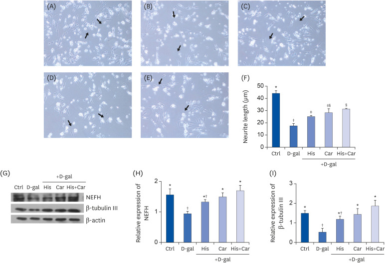
Fig. 2. Effects of L-histidine, L-carnosine, and their combination on neuronal cell regenerations.
Representative pictures for each group (100× magnification). (A) Ctrl, (B) D-gal, (C) His, (D) Car, and (E) His+Car. (F) The average length of the neurites of differentiated SH-SY5Y cells for each group was analyzed. ImageJ software was used to measure the individual neurite length. The protein expressions of an axonal marker, NEFH, and a neuronal marker, β-tubulin III were determined by Western blotting and β-actin was used as a loading control. (G) Representative blots are shown. Quantification of NEFH (H) and β-tubulin III (I) levels to β-actin are shown. The values shown are the mean ± standard error of the mean (n = 3–4).
NEFH, Neurofilament heavy polypeptide; Ctrl, Control; D-gal, 200 mM D-galactose; His, 1 mM L-histidine; Car, 10 mM L-carnosine; His+Car, 1 mM L-histidine + 10 mM L-carnosine.
*,†,‡,§Different superscript marks indicate significant differences between groups (P < 0.05).

