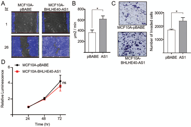
Figure 3. BHLHE40-AS1 supports an invasive phenotype.

Panel A: 2.0x10⁵ MCF10A-pBABE and MCF10A-BHLHE40-AS1 cells were plated in 6 well dishes. Confluent cells were scratched with a pipette tip 72h after seeding, rinsed with PBS and replenished with fresh medium. Cells were imaged every 30 min to monitor wound closure. Representative examples of the scratch assay are shown. Panel B: Rate of wound closure. Open area was calculated using ibidi MetaViLabs Automated Cellular Analysis System. Bars represent mean ± SD of 3 independent biological replicates * p<0.05, two-tailed Student’s t test. Panel C: 2.0x10⁵ MCF10A-pBABE and MCF10A-BHLHE40-AS1 cells were plated in 6 well dishes. 48h post plating, cells were evaluated for invasive potential by a Boyden chamber assay. 2.5 x 10⁴ cells were added to the upper chamber in serum free media and cultured for 24h. Cells were fixed with methanol, stained with crystal violet and counted using ImageJ. Bars represent mean ± SD of 3 independent biological replicates * p<0.05, two-tailed Student’s t test. Panel D: 4 x 10³ cells were seeded into 96 well plates and cell number was measured using the CTG assay at the indicated time points.
