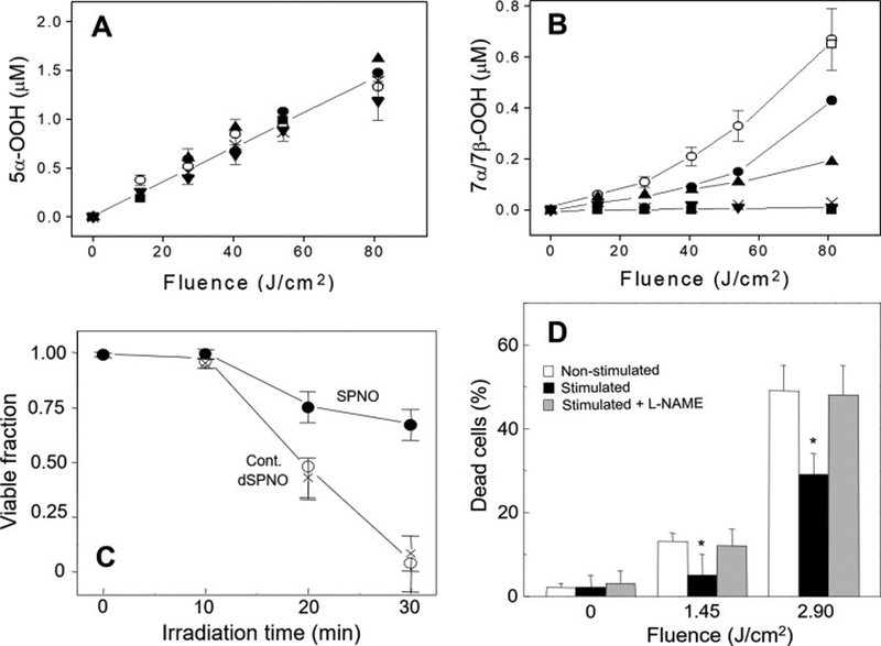
Figure 1.

Effect of NO on photosensitized ChOOH formation in liposomes and on necrotic killing of cancer cells. (A) Accumulation of 5α-OOH and (B) 7α/7β-OOH in POPC/Ch/PpIX (100:80:0.2 by mol) liposomes irradiated in the absence vs. presence of 1 mM ascorbate (AH), 0.5 μM ferric-8-hydroxyquinoline (Fe) and spermine-NONOate (SPNO): −Fe/AH (x); +Fe/AH (○); + Fe/AH and 20 μM SPNO (●), 60 μM SPNO (▲), 200 μM SPNO (▼), 400 μM SPNO (■), or decomposed 400 μM SPNO (□). ChOOHs in lipid extracts were determined by HPLC-EC(Hg). (C) Photokilling of disseminated PpIX-sensitized COH-BR1 cells in the absence (○) vs. presence of 400 μM SPNO (●) or decomposed SPNO (x). Light fluence at 30 min irradiation: 2.2 J/cm2. (D) Photokilling of COH-BR1 cells during exposure to non-stimulated or stimulated RAW 264.7 macrophages (15 h after LPS added) in absence vs. presence of 4 mM L-NAME. All values are means ± SEM (n=3). *P<0.005 vs. non-stimulated ± L-NAME. (Reproduced from Refs. 18 and 19, with permission.)
