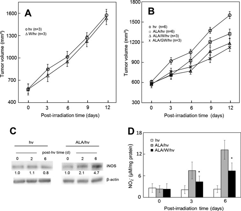
Figure 3.

Antagonistic effects of iNOS/NO on ALA-PDT in a tumor xenograft model. (A) Light-only controls: female SCID mice bearing human breast MDA-MB-231 tumors were injected i.p. with PBS or 1400W (10 mg/kg) in PBS; after 4 h, the mice were anesthetized, placed in opaque restraints with cutouts for tumor exposure, and irradiated using a 633-nm light source and fluence ~95 J/cm2; after irradiation, mice were reinjected with 1400W once per day until termination; plotted tumor volumes are means ± SEM (n=3). (B) ALA-PDT: Tumor-bearing mice were injected i.p. with ALA (100 mg/kg) or PBS (light-only controls), followed by 1400W (10mg/kg) or GW274150 (25 mg/kg); after 4 h in the dark, the mice were anesthetized, placed in restraints with cutouts, and irradiated as in (A); after PDT, mice were kept in subdued light and reinjected with each iNOS inhibitor once daily; measured tumor volumes are means ± SEM (n=3–6). (C) Western blots showing iNOS levels in tumor samples from light control and ALA-PDT mice; number below each band indicates iNOS intensity relative to β-actin and normalized to day-0. (D) Nitrite levels in tumor samples after ALA-PDT in the absence vs. presence of 1400W; values determined by Griess assay are means ± SEM (n=3). (Reproduced from Ref. 41, with permission.)
