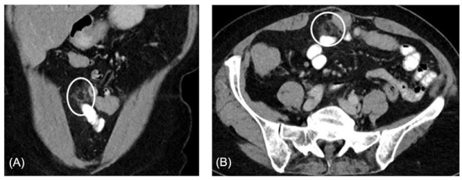
Figure 1.

(A), Example of contrast enhanced longitudinal abdominal CT demonstrating a primary epiploic appendagitis adjacent to the transversal colon in a 68-year-old white female patient. The lesion, with a size of size 2.3 x 1.2cm, shows fat attenuation and surrounding inflammation. (B), In the same patient an axial abdominal CT with contrast enhancement showing primary epiploic appendagitis adjacent to the transversal colon in. The lesion demonstrates fat attenuation and surrounding inflammation.
