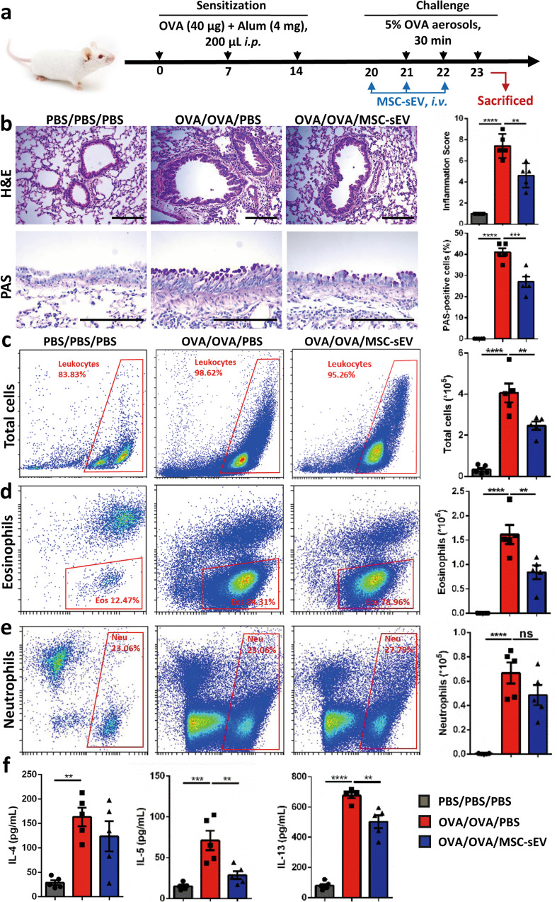
Fig. 2. The effects of iPSC-MSC-EVs in eosinophilic allergic airway inflammation.
a Schematic diagram for the development eosinophilic airway inflammation in mice and therapy interventions of MSC-sEV. b Representative figures and statistical results of H&E and PAS staining for lung tissues. c–e Cell counts for inflammatory cells by flow cytometry in BALF. f Levels of Th2-related cytokines in the BALF. **P < 0.01, ***P < 0.001, ****P < 0.0001. Alum Aluminum, BALF bronchoalveolar lavage fluids, EVs Extracellular Vesicles, H&E Hematoxylin–Eosin, i.p. intraperitoneally, MSC Mesenchymal stromal cells, ns not significant, OVA ovalbumin, PAS periodic acid-schiff, PBS phosphate buffer saline. N = 5 for PBS/PBS/PBS, OVA/OVA/PBS and OVA/OVA/MSC-EVs. Scale bar, 150 µm.

