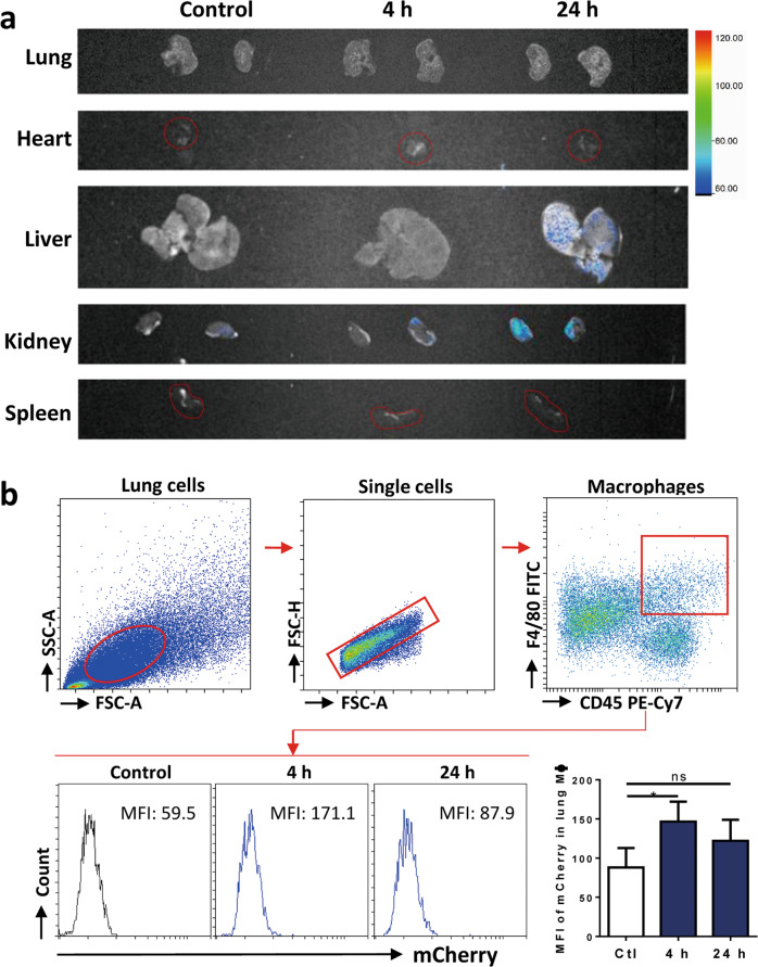
Fig. 4. Macrophages were able to take MSC-sEV in both in vitro and in vivo.
a Mice were administered with mCherry-labeled iPSC-MSC-sEV, and whole-body imaging was performed to detect the distribution of sEV at the indicated time points. b Total lung cells were isolated and MFI of mCherry in macrophages was analyzed by means of flow cytometry. Pulmonary macrophages significantly uptook MSC-EVs at 4 h post-administration. *P < 0.05. MFI mean fluorescence intensity, ns not significant.

