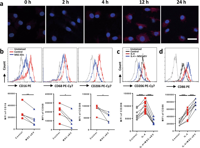
Fig. 5. The effects of MSC-sEV on the differentiation and polarization of human Mo–Mϕ in vitro.
Macrophages were induced from human CD14+ monocytes. a Macrophages were treated with mCherry-labelled MSC-sEV (red) for different times. The nuclei were counterstained with 4′,6-Diamidine-2′-phenylindole dihydrochloride (blue). b CD14+ monocytes were cultured with M-CSF for 7 days to differentiate into macrophages, and the expression of CD16, CD68 and CD206 on macrophages were determined by flow-cytometry analysis. c, d Mature macrophages were polarized into M2 and the expression of CD206 (M2 marker) and CD86 (M1 marker) was determined by flow-cytometry analysis. *P < 0.05, **P < 0.01, ***P < 0.001. MSCs mesenchymal stromal cells, MFI mean fluorescence intensity, sEV small extracellular vesicles. Scale bar, 20 µm.

