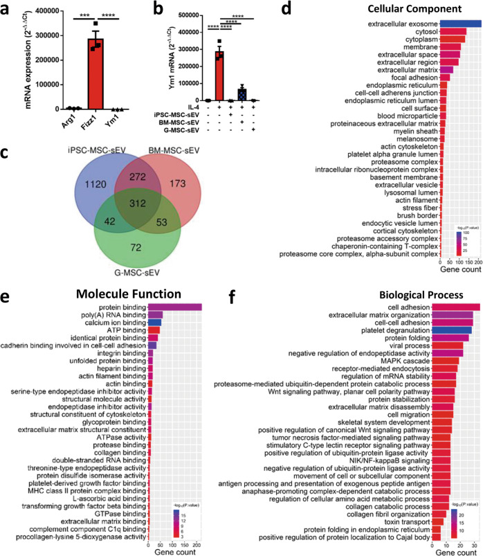
Fig. 6. Proteomic signatures of MSC-sEV.
a, b Raw 264.7 macrophages were stimulated with IL-4 for 48 h for polarization of M2 macrophages (a), and were treated with three types of MSC-sEV. The mRNA levels of Arg1, Fizz1, and Ym1 were determined by qRT-PCR (b). c Venn diagram for the numbers of protein identified by iPSC-MSC-sEV, BM-MSC-sEV, and G-MSC-sEV. d–f GO analysis of the protein co-expressed by iPSC-MSC-sEV, BM-MSC-sEV, and G-MSC-sEV. ***P < 0.001 and ****P < 0.0001. BM-MSCs bone marrow-derived mesenchymal stromal cells, G-MSCs gingiva-derived mesenchymal stromal cells, iPSC-MSCs induced pluripotent stem cell-derived mesenchymal stromal cells, sEV small extracellular vesicles.

