Figure 1.
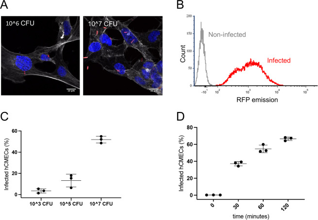
Infection by E. coli EV36-RFP of hCMEC cultures. (A) Representative fluorescent images showing E. coli EV36-RFP infection of hCMEC cultures at concentrations of 106 CFU/ml (left) and 107 CFU/ml (right). E. coli EV36-RFP fluorescence shown in red, DAPI in blue, and phalloidin in white (B) Flow cytometry histogram showing E. coli EV36-RFP infected hCMECs in red and non-infected hCMECs in grey, as detected by RFP fluorescence. (C,D) Mean percentages of E. coli EV36-RFP infected hCMECs (RFP-positive by flow cytometry) after a 1 h incubation period with concentrations of 103 to 107 CFU/ml (C), or 107 CFU/ml sampled over time (D); + /− SD, n = 3 in each case.

