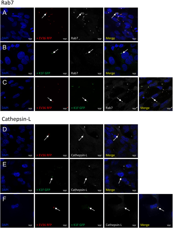
Figure 3.
Lysosomal- and phagosomal markers of constitutive phagocytosis. (A–F) Immunofluorescent images showing hCMEC cultures fixed and stained with anti-RAB7 (A–C) and anti-Cathepsin-L (D–F) antibodies following a 1 h incubation with 107 CFU/ml E. coli EV36-RFP alone (A + D) or 107 PFU/ml phage K1F-GFP alone (B + E), or a 1 h incubation with 107 CFU/ml E. coli EV36-RFP followed by a 1 h incubation with 104 PFU/ml phage K1F-GFP (C + F). DAPI stain is shown in blue and anti-RAB7/anti-Cathepsin-L antibodies in white. n = 3 in each case.

