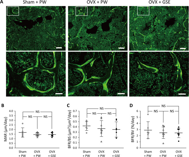
Figure 5.
Histological and histomorphometric evaluation of osteogenic parameters in the trabecular bone in ovariectomized (OVX) mice administered with proanthocyanidin-rich grape seed extract (GSE). (A) Representative fluorescent microscopy images showing the calcein (green) labels of the trabecular bone sections of lumbar vertebrae from each of the three groups. The area under the white square in the top row was analyzed at higher magnification as shown in the bottom row. Scale bars in the top and bottom row represent 200 and 50 µm, respectively. (B–D) Quantification of osteogenesis-associated histological measures. In (B–D), the results are expressed as the mean ± standard deviation, showing individual data (n = 6 in Sham + PW, n = 8 in OVX + PW, n = 7 in OVX + GSE). MAR: mineral apposition rate, BFR/BS: bone formation rate/bone surface, and BFR/BV: BFR/bone volume. No significant differences in any of the three measures were found between the three groups.

