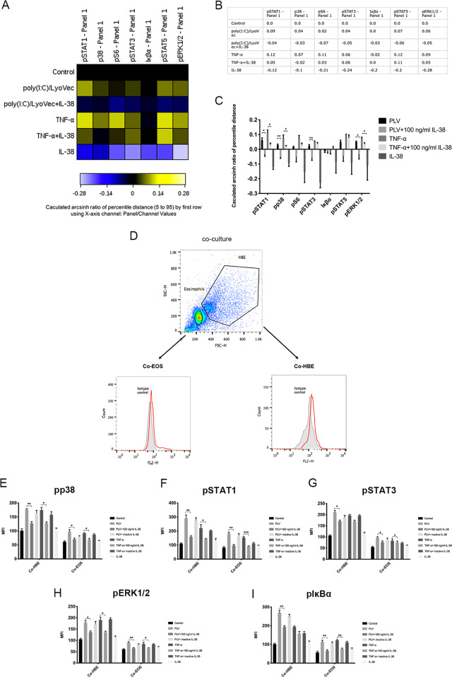
Fig. 3.
Mass cytometric profiling of intracellular signaling pathways. Cocultured cells were pretreated with human IL-38 (100 ng/ml) for 10 min and then stimulated with poly (I:C)/LyoVec (2 μg/ml) or human TNF-α (20 ng/ml) for an additional 20 min. The phosphorylated signaling molecules in the stimulated cells were stained by using the Maxpar® Phospho Panel Kit. a A heatmap was generated by Cytobank. b, c The change in each phosphoepitope (calculated as the arcsinh difference of the 95th percentile) was quantified for analysis. d Gating strategies for bronchial epithelial cells and eosinophils from cocultures are shown. The intracellular levels of e phosphorylated p38, f phosphorylated STAT1, g phosphorylated STAT3, h phosphorylated ERK1/2, and i phosphorylated IκBα in cells were measured by intracellular staining with specific antibodies and analyzed by flow cytometry. The absolute MFI values for the phosphorylated signals were used for analysis. Heat-inactivated human IL-38 (100 ng/ml) served as a negative control. The results are shown as the mean ± SEM of triplicate independent experiments with a total of three donors. Abbreviations: Co-HBE, human primary bronchial epithelial cells in coculture; Co-EOS, eosinophils in coculture *P < 0.05, **P < 0.01, and ***P < 0.001 when compared between the denoted groups

