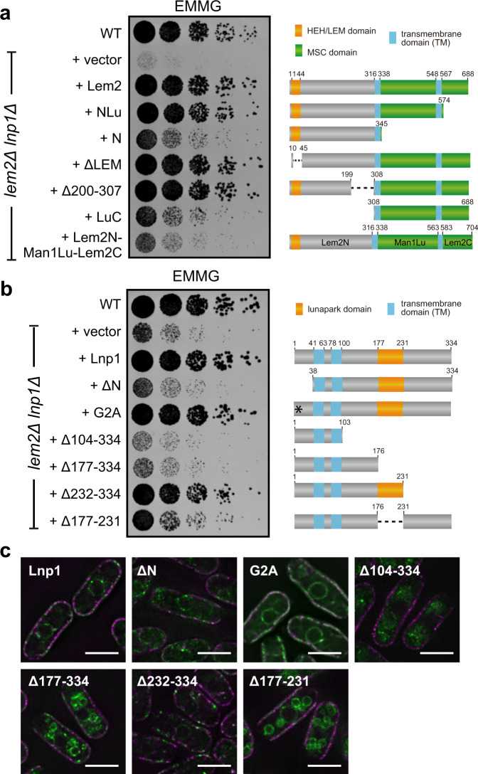
Fig. 4. Functional domains of Lem2 and Lnp1.
a Lem2 fragments indicated on the left were expressed in lem2Δlnp1Δ cells. Diagram of these fragments is shown on the right. Fivefold serially diluted cells were spotted on EMMG plates, and growth of these cells was observed after 3 days. b Lnp1 fragments indicated on the left were expressed in lem2Δlnp1Δ cells. Diagram of these fragments is shown on the right. The asterisk indicates the mutation site of G2A. Fivefold serially diluted cells were spotted on EMMG plates, and growth of these cells was observed after 3 days. c Localization of Lnp1 fragments GFP-tagged Lnp1 fragments (green) and cortical ER membrane marker Rtn1-mRFP (magenta) were expressed in wild-type cells. Scale bar represents 5 μm.

