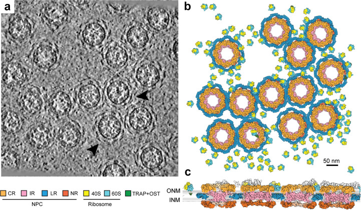
Fig. 1. Three-dimensional organization of the NPCs in a local region of the X. laevis NE.
a Organization of the NPCs in an original tomogram slice as viewed along the nucleocytoplasmic axis. Some of the representative array-like densities of the LR are indicated by arrowheads. The thickness of the tomographic slice shown here is 8.9 Å. b Organization of the NPCs in the reconstructed tomogram as viewed along the nucleocytoplasmic axis. As the outer boundary of the NPC, the LR appears to cushion the contacts among neighboring NPC particles. Reconstructions for the individual NPC subunits (CR, IR, NR and LR) and the ribosomes associated with TRAP + OST46 were back-projected onto the original tomograms based on the refined coordinates of the individual particles. Shown here is a section of the NE from a. Scale bar, 50 nm. c Organization of the NPCs in the reconstructed tomogram as viewed perpendicular to the nucleocytoplasmic axis. In contrast to other ring scaffolds of the NPC, the LR resides in the lumen. 40S: Small ribosome subunit; 60S: Large ribosome subunit; TRAP: translocon-associated protein complex; OST: oligosaccharyl-transferase.

