Figure 1.
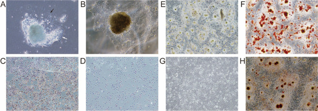
OSCC cell features. (A) OSCC cells (black arrow) and fibroblasts (white arrow) direct outgrowth from the OSCC specimens. (B) OSCCs after a 15-day culture. (C) Brown staining was positive in the cytoplasm of OSCC cells detected by immunohistochemical staining for keratin. (D) Minimal brown staining was observed in the cytoplasm of OSCC cells in the blank control group. (E,F) CD133+ OSCC cells formed in adipose tissue observed by microscopy following Oil Red O staining. (G,H) Calcified nodules formed by CD133+ OSCC cells stained with Alizarin Red.

