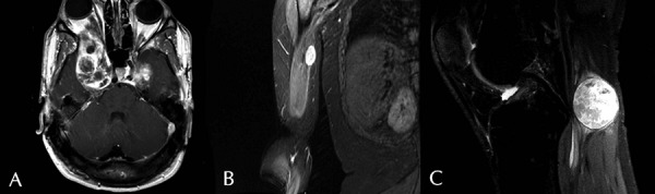
Figure 1. Post-contrast T1 weighted MR image. A: Axial image displaying large enhancing tumors of the cavernous sinus and right orbit (Patient 11). B: Coronal image of right arm showing enhancing tumor (Patient 4). C: Sagittal image of right knee depicting enhancing tumor on right tibial nerve (Patient 3).