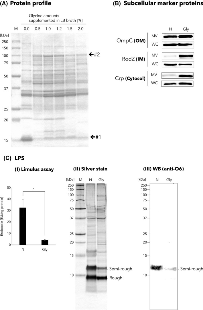
Fig. 4.

Analysis of the amounts of subcellular marker proteins and LPS.
A. Protein profiles of MVs isolated in different culture conditions, in which the LB broth contained different amounts of glycine (0–2.0%). The same amount of protein (1.7 µg) was loaded into each lane of the SDS‐PAGE. Locations of bands #1 and #2 in the CBB‐stained SDS‐PAGE gel are indicated. See also Table 2, in which the results of protein identification of these bands are shown. ‘M’ represents marker.
B. Analysis of subcellular marker proteins. Western blotting analyses of OmpC, RodZ and Crp proteins in MV and whole‐cell (WC) samples prepared from LB media supplemented without (N) or 1.0% glycine (Gly). The same amount of protein was loaded into each lane.
C. LPS analyses of MVs. (I) Limulus assay. The amount of LPS in glycine‐induced and non‐induced MVs. Shown are the mean values with standard errors of the endotoxin unit (EU) following standardization with the protein amounts (ng). Statistical analysis was performed using unpaired t‐test with Welch's correction (n = 3, *P < 0.05). (II) Silver stain. Protein profiles of non‐induced and glycine‐induced MVs of EcNΔflhD were assessed by silver staining. The same amount of protein was loaded into each lane. MV samples were separated by Tris‐Tricine SDS‐PAGE using 16.5% polyacrylamide gels and stained with silver nitrate. (III) WB [anti‐O6]. The same amount of protein was loaded into each lane. MV samples were separated by Tris‐Tricine SDS‐PAGE using 16.5% polyacrylamide gels. LPS O6 antigen in the MV samples was probed with E. coli O6 antiserum. A single nucleotide mutation is located in the wzy gene (encodes O‐antigen polymerase) of the O6 antigen synthesis gene cluster in the chromosome of the EcN strain (Grozdanov et al., 2002). Due to the single nucleotide exchange in the wzy gene, EcN LPS lacks long O‐antigen. Shown are two different LPS species, a semi‐rough‐type (Semi‐rough) and rough‐type (Rough), composed of lipid A‐core oligosaccharide with and without a single O‐antigen repeating unit respectively (Grozdanov et al., 2002). Hence, only semi‐rough species that contain one unit of O6 oligosaccharide are detectable with anti‐O6 antiserum.
