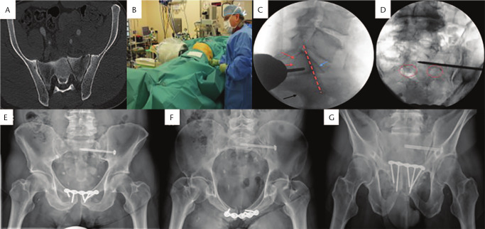
Fig. 4.
(A) Axial CT scan showing Dennis I fracture over left sacral ala; (B) Patient in supine position, under fluoroscopic control and left buttock stab incision, surgeon tries to identify correct position of guide wire for sacral body 1 screw insertion; (C) Lateral fluoroscopic image demonstrating upper end of the first sacral vertebra (blue arrow), the iliac cortical density (red line), the greater sciatic notch (black arrow) and the upper nerve root tunnel (red arrows); (D) S1 sacral foramina are demonstrated by red circles. Guide wire for screw insertion is positioned above left S1 foramen; (E) Postoperative anteroposterior (AP) pelvic radiograph; (F) inlet and (G) outlet views demonstrating fixation of the pelvic ring (injury to pubis symphysis was addressed with a plate).

