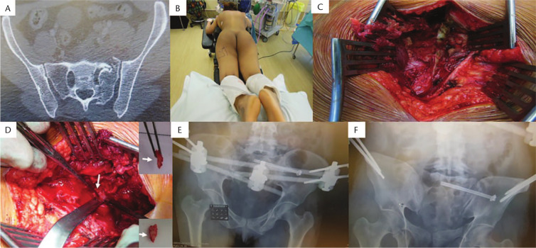
Fig. 5.
(A) Axial computed tomography (CT) pelvic slice showing Denis II fracture with a small bone fragment in the Sacral 1 (S1) foramen causing S1 nerve root compression. (B) Patient positioned in prone position. (C) Midline exposure and for decompression of the nerve root. (D) Bony fragment removal (arrows). (E and F) Pelvic ring fracture was fixed with left SI sacroiliac screw and an anterior external fixator frame for the pubic rami fracture.

