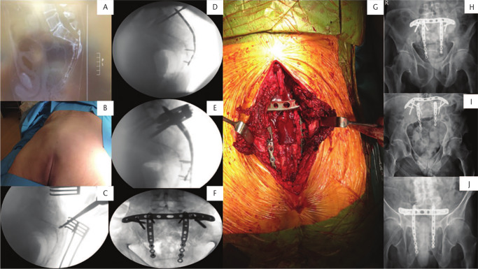
Fig. 7.
(A) Lateral sacral radiograph showing a U-type fracture. Patient presented with full sacral plexus neurological symptoms, incontinence. Underwent diverting stoma post decompression of spinal canal and orif with tension band plate and bilateral sacral wing locking plates to address vertical fracture plane at levels of S1–S3. (B) Prone position of patient. (C) Fluoroscopic image for identification of the fracture level and decompression. (D) Lateral fluoroscopic image showing application of sacral wing plates and insertion of sacroiliac (SI) body screw. (E) Lateral fluoroscopic view showing fixation of sacral fracture with tension band plate and bilateral sacral wing locking plates. (F) Fluoroscopic outlet view showing fixation of fracture. (G) Intraoperative picture showing midline incision and application of plates. (H) Anteroposterior, (I) inlet, and (J) outlet pelvic postoperative radiographs showing stabilization of the ring posteriorly.

